CORE A
IMAGE
Here is an image of Core A. It is a long core section, so scroll down. The clear round pins on the left indicate the depth from the top of the core marked every 10 cm (ex: 800 = 800 cm down from the sediment surface).

MICROSCOPE IMAGE
To determine the sediment type, a slide was made of a small sample of sediment and examined under the microscope. Here is what the “smear slide” looks like under special polarizing light.
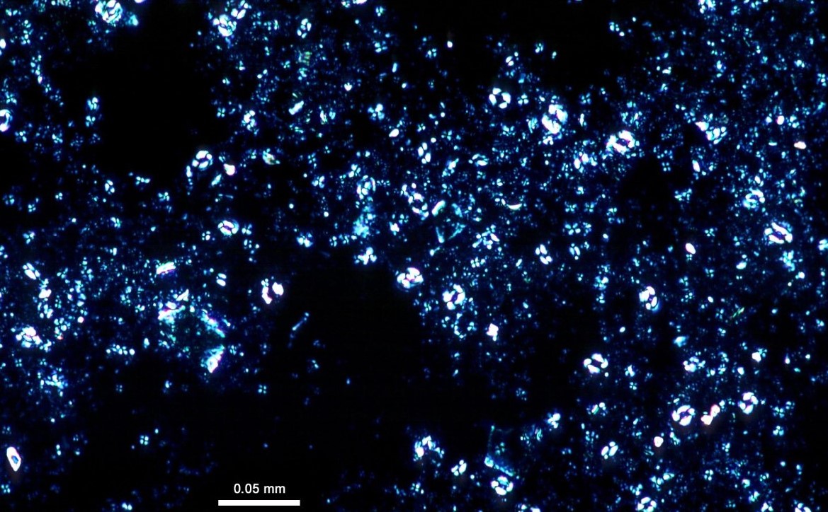
Image source: https://tmi.laccore.umn.edu/is
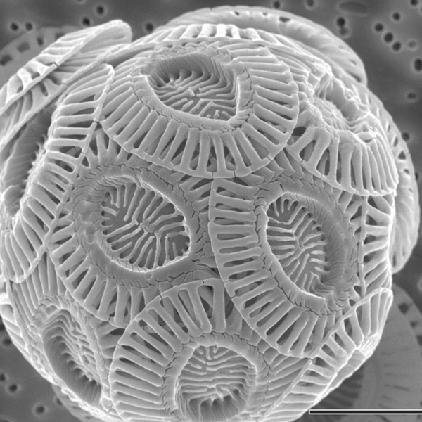
The sediment is so small that it would take a scanning electron microscope to actually see the particles. Below is an image from a similar sediment sample.
SMEAR SLIDE ANALYSIS
A close analysis of the slide provided the following components:
Calcareous nannofosssils: 95%
Foraminifera: 2%
Diatoms: 1%
Radiolaria: 2%