CORE B
IMAGE
Here is an image of Core B. The scale bar on the left indicates depth of sediment in cm.

CT SCAN
CT scans are also useful tools to look at cores. Like an xray, dense material appears whiter, while holes and cracks in the core appear black. The CT scan of the upper part of Core B is shown below. Note the distinct layers that are more clearly visible in the CT scan compared to the camera image.

MICROSCOPE IMAGE
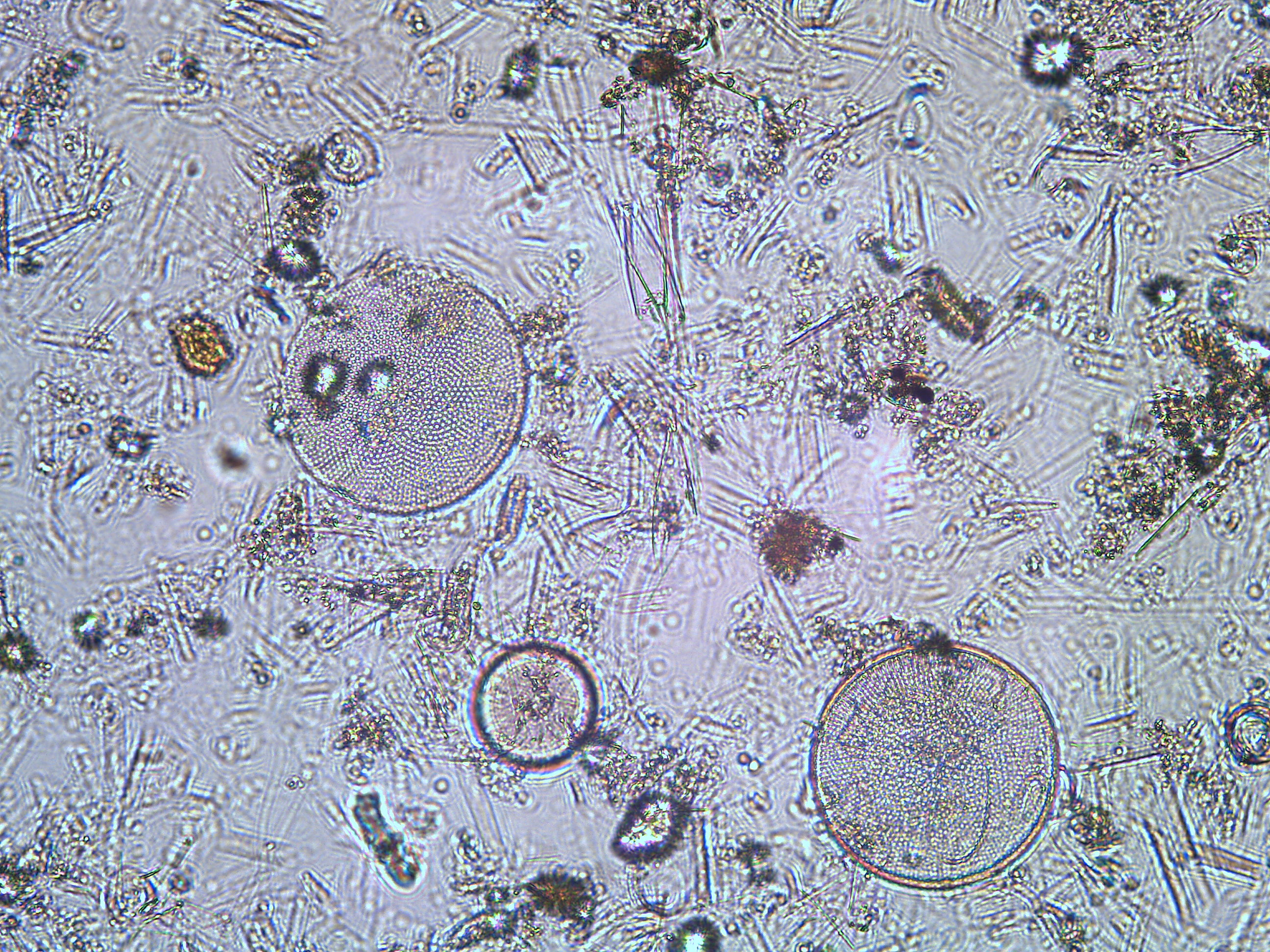
Let’s look closer at what one of these layers is composed of. Here is a microscope image of sediment from one of these layers.

SMEAR SLIDE ANALYSIS
A close analysis of the slide provided the following components:
Sponge spicules: 75%
Diatoms: 20%
Clay minerals: 5%
